
Polychromasia
Hyperchromia
Cytochemical Staining of Leukemias
Stain |Site of Action |Cells Stained |Comments |Image
Myeloperoxidase
Primary Granules;
Auer Rods
Myeloblasts; Granulocytes;
Monocytes slight positive
Separates AML+ from AML-
Sudan Black B Phospholipids
Myeloblasts; Granulocytes;
Monocytes slight positive
Separates AML+ from AML-
Naphthol AS-DChloroacetate/(CAE) – specific esterase
Cytoplasm
Neutrophilic granulocytes;
Mast cells
Separates AML+ from AML-
Periodic acid-Schiff Stain (PAS)
Glycogen Granular pattern with negative background in lymphoblasts.
Positive in erythroleukemia, abnormal erythrocyte precursors, and ALL
Also, histochemical staining for terminal deoxynucleotidyl transferase (TdT) can be useful in determining that the blast is of lymphoid lineage. TdT is an enzyme found in immature (developing) lymphocytes which functions to help synthesize their specific antigen receptors.
Chronic Myeloid Leukemia
BCR-ABL test for Philadelphia chromosome: A gene formed when pieces of chromosomes 9 and 22 break off and trade places.
t (8;21) – Acute Myeloid Leukemia (FAB M2)t (8;14) – Burkitt’s Lymphomat (11;14) – Chronic Lymphoid Leukemia and multiple myeloma
Kohler Illumination and Microscope Calibration

Accute ProMyelocytic Leukemia (APML)
Start ATRA STAT
B cell Lymphoma
AKA Hairy Cell lymphoma
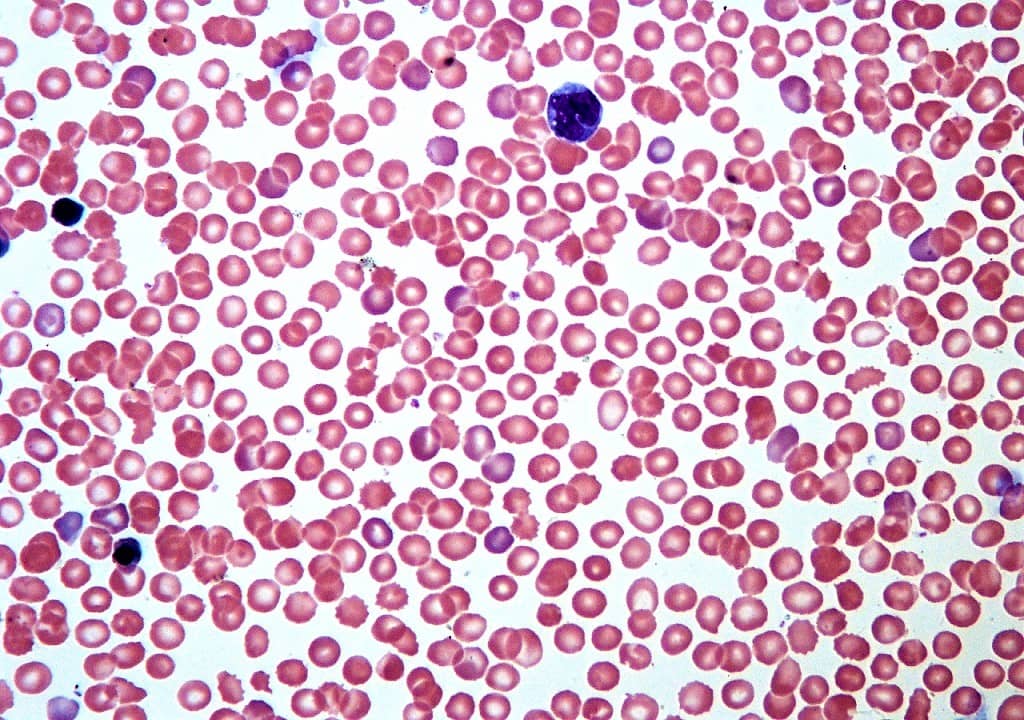
CBC and Auto-Differential
Hemacytometers were the first tools used to perform manual cell counts. A standard ‘charge’ would fill a 3×3 gridded area which was eactly 0.9uL. The coulter counter was one of the first automated laboratory instruments and would count using a tube so skinny exactly one cell could fit through its diameter. A constant electrical charge would be flowing across a part of the cross section and the charge would be impeded by a cell traveling through through. The resistance was measured and directly correlated to cellular volume.
MCHC = HGB * 100 / HCT
MCH = HGB *10 / RBC
MCV = HCT * 10 / RBC
A Word on Motivation
https://www.dataquest.io/blog/does-sharing-goals-help-or-hurt-your-chances-of-success/